
|
By Tom Volk
Besides being interesting for its edibility and controversial taxonomy, this month's fungus has lots of history behind it. Amanita caesarea and its American relatives Amanita hemibapha and Amanita jacksonii are among the relatively few widely-consumed edible Amanita species. The genus Amanita is better known for its poisonous members the death angels (Amanita virosa, A. bisporigera, A. verna), the destroying angel or death cap (A. phalloides) and the hallucinogenic and toxic fly agaric (A. muscaria). There are many hundreds of species of Amanita across all the continents (except Antarctica), including dozens if not hundreds of species that are still unknown to science, waiting to be discovered. The species of Amanita are fairly narrowly defined, and we have little information about the edibility of most species. Because of the possibility of poisoning combined with the difficulty of identifying the species correctly, you should be very careful about eating any Amanita specimen. Amanita is definitely not recommended for mycological beginners. Every year many people are poisoned, thinking they are eating an edible species when they are in fact eating a deadly Amanita. Amanita is a well-defined genus of mycorrhizal Agaricales (gill forming mushrooms) that have a white spore print, gills (lamellae) that are free from the stipe (stalk) and a universal veil covering the young mushroom buttons. When the mushroom expands, the universal veil is broken; the bottom of the universal veil forms the cup shaped volva at the base of the mature mushroom. The top of the universal veil is often left at the top of the mushroom cap, forming a patch or sometimes breaking up into scaly floccules. Most species also have a partial veil, a membranous structure that protects the developing gills of the young mushroom. When the mushroom cap expands, the partial veil breaks and is left as a ring (annulus) on the stalk. See the picture to the right for labeling of these various parts of the mature mushrooms and the expanding button. Some Amanita species, such as A. fulva and A. vaginata lack the partial veil and annulus. Almost all other genera of mushrooms lack the universal veil and volva-- a notable exception is Volvariella, but that genus has a pink spore print and always grows on wood or other debris, including other mushrooms. As you might guess from the name, Amanita caesarea was a favorite of the emperors of the Roman Empire, the Caesars. Perhaps Julius Caesar ate a meal of this delicious mushroom before Brutus did him in on the Ides of March (March 15). Et tu Brute? However, the most famous killing involving Amanita took place in ancient Rome circa 50-60 A.D. The Emperor Claudius had ascended to the throne after the assassination of his nephew Caligula. We've all heard about the decadence (and Roman porno movies, such as "Veni, Vidi, Veni") associated with Caligula. Anyway, Claudius had several wives, but finally married his fourth wife Agrippina, who was also his niece. Agrippina already had a son from a previous marriage, Nero, for whom she had great plans. She persuaded Claudius to adopt Nero, putting him in line for the throne, should something happen to Claudius. (You can see where this is going already.) Agrippina was an impatient woman, and could not wait for a natural death for Claudius; she plotted to kill him by feeding Claudius his favorite meal, Amanita caesarea, laced with extracts from Amanita phalloides, the death cap. When the symptoms set in the next day, a co-conspirator doctor Xenophon administered an enema of colocynth, a potent toxin from a plant called bitter apple, which together with the mushroom toxin killed Claudius. Nero and his fiddle thus became emperor, and the rest, as they say, is history. It is pretty well accepted that true Amanita caesarea does not exist in North America, having been described from the Italian region. So although Amanita caesarea is the fungus of the month, none of the picture on this page are really Amanita caesarea. To the right is (probably) Amanita hemibapha from Texas; the pictures above, from Connecticut, are likely Amanita jacksonii. These two species are collective called the "American Caesar's Mushroom." As currently circumscribed, Amanita species are very narrowly defined, and species identification is difficult, at best. There are significant microscopic differences between the three species, and until someone does the molecular (DNA) studies on this group, we should consider them to be different species. The picture to the right also illustrates why you should carefully dig under mushrooms when you are collecting them. If you just pulled these mushrooms out by their stems you might miss the diagnostic character, the volva. Looking back at the pictures on this page, I should also point out that these mushrooms are *much* more beautiful when you see live fresh specimens. There are also much better, very beautiful pictures of these mushrooms in existence, but I do not see this mushroom very often-- never in Wisconsin. 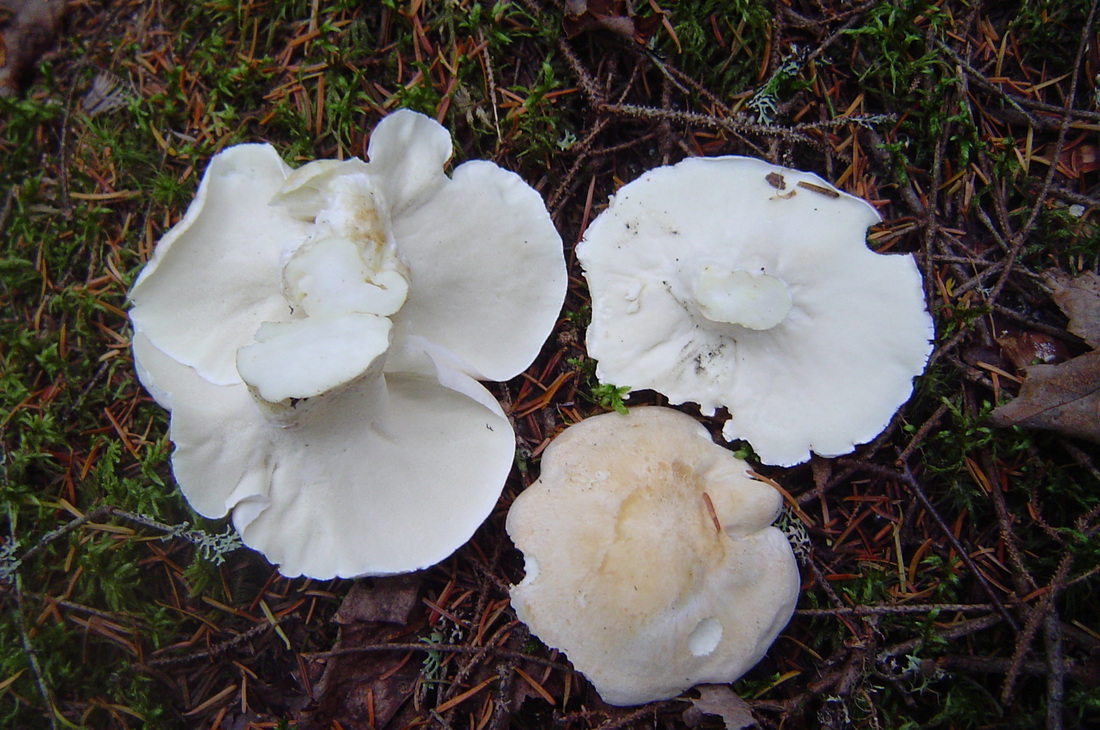 location: North America, Europeedibility: Ediblefungus
colour: White to cream, Grey to beige normal size: 5-15cm cap type: Convex to shield shaped stem type: Simple stem flesh: Pore material cannot be seperated from flesh of the cap spore colour: White, cream or yellowish habitat: Grows in woods, Grows on the ground Sheep Polypore Albatrellus ovinus (Fr.) Murr. syn. Polyporus ovinus Fr. Schafporling Fakó zsemlegomba. Fruit body annual. Cap 5-15cm across, usually single but sometimes several fused together, circular to irregular when fused, convex then depressed, dish-shaped; white to pale buff, tan; dry, smooth, or a little scaly with age. Tubes 1-2mm deep, decurrent; white. Pores 2-4 per mm, angular; white to yellowish. Stem 20-75 x 10-30mm, slightly swollen, pointed at base, usually central; white bruising pinkish; smooth. Flesh 5-20mm thick, firm; white, dries yellowish. Odor pleasant, fungusy, aromatic. Taste mild, sometimes slightly bitter. Spores subglobose-ellipsoid, 3-4.5 x 3-3.5µ. Deposit white. Hyphal structure monomitic. Habitat on the ground by conifers, especially at high elevations. Found in Europe especially Finland (where it is considered a fine edible,)and throughout North America. Season August to winter. Edible. Comment Similar are Albatrellus confluens (Fr.) Kotlaba & Pouz., which is darker, orange-hued, with a bitter flavor, and Albatrellus subrubescens (Murr.) Pouz., which bruises orange. http://www.rogersmushrooms.com/gallery/DisplayBlock~bid~5514.asp 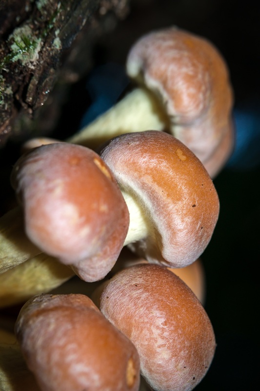 by Michael Kuo
This widely distributed fall mushroom, sometimes called the "brick cap," can be found growing in tight clusters on hardwood stumps and logs. It is fairly easily recognized by its habitat, its brick-red cap with a paler cap margin, its purple-gray gills, and the way the stem often bruises and stains yellow. If you can catch Hypholoma sublateritium when it's still very young, you can see its partial veil, which mycologists call "submembranous," looking like a cross between a cortina and a more substantial veil. Description: Ecology: Saprobic; growing in clusters on decaying hardwood logs and stumps; fall; widely distributed in North America but more common east of the Rocky Mountains. The illustrated and described collections are from Michigan, Illinois, and Québec. Cap: 3-10 cm; convex, becoming broadly convex, nearly flat, or irregular in age; with an incurved margin when young; bald; dry or moist; brick red overall, but paler (pinkish to buff) on the margin, especially when young; the margin sometimes hung with wispy veil fragments. Gills: Attached to the stem; close or crowded; when young covered by a whitish, cortina-like veil; whitish when very young, but soon pale gray to gray, becoming purple-gray to dark purple brown with maturity; short-gills frequent. Stem: 4-12 cm long; 1-2 cm thick; more or less equal, or twisted and tapering to base due to the clustered growth pattern; bald, or finely hairy near the apex; often featuring an ephemeral or persistent ring zone near the top; yellowish to whitish above, brown to reddish below; sometimes bruising and staining yellow. Flesh: Firm; whitish to yellowish. Odor and Taste: Odor not distinctive; taste mild or slightly bitter. Chemical Reactions: KOH brownish on cap surface. Spore Print: Purple brown. Microscopic Features: Spores 6-7 x 3-4 µ; ellipsoid; smooth; thin-walled; with an obscure pore; yellowish in KOH. Pleuro-chrysocystidia abundant; fusoid-ventricose to mucronate; up to 40 x 10 µ. Cheilocystidia fusoid-ventricose, cylindric, or irregular, with subcapitate apices; hyaline; thin-walled; to 35 x 8 µ. Pileipellis a cutis; elements golden in KOH. REFERENCES: (Schaeffer, 1774) Kummer, 1871. (Saccardo, 1887; Smith, 1949; Smith, 1951; Stamets, 1978; Smith, Smith & Weber, 1979; Phillips, 1991/2005; Lincoff, 1992; Barron, 1999; Roody, 2003; McNeil, 2006; Miller & Miller, 2006.) Herb. Kuo 09059508, 11060402, 11290901. Naematoloma sublateritium is a synonym. European mycologists often list Hypholoma sublateritium as a deprecated synonym of Hypholoma lateritium. Further Online Information: Hypholoma sublateritium at Roger's Mushrooms
12/1/2015
December 2015 Psilocybe cubensis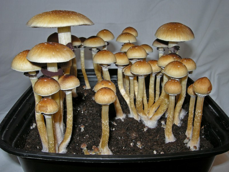 Psilocybe cubensis is a species of psychedelic mushroom whose principal active compounds are psilocybin and psilocin. Commonly called shrooms, magic mushrooms, golden tops, cubes, or gold caps, it belongs to the Hymenogastraceae family of fungi and was previously known as Stropharia cubensis. It is the most well known psilocybin mushroom due to its wide distribution and ease of cultivation. The species was first described in 1906 as Stropharia cubensis by Franklin Sumner Earle in Cuba.[1] In 1907 it was identified as Naematoloma caerulescens in Tonkin by Narcisse Théophile Patouillard,[2] while in 1941 it was called Stropharia cyanescens by William Alphonso Murrill in Florida.[3] These synonyms were later assigned to the species Psilocybe cubensis.[4][5]
The name Psilocybe is derived from the Greek roots psilos (ψιλος) and kubê (κυβη),[6] and translates as "bald head". Cubensis means "coming from Cuba", and refers to the type locality published by Earle. Psilocybe cubensis Pileus: 2–8 cm, Conic to convex, becoming broadly convex to plane in age, may retain a slight umbo, margin even, reddish-cinnamon brown when young becoming golden brown in age, viscid when moist, hygrophanous, glabrous, sometimes with white universal veil remnants decorating the cap, more or less smooth. Flesh whitish, bruising blue in age or where injured. Gills: Adnate to adnexed to sometimes seceding attachment, close, narrow to slightly wider towards the center, at first pallid to gray, becoming dark purplish to blackish in age, somewhat mottled, edges remaining whitish. Spore Print: Dark purple brown Stipe: 4–15 cm long, .5–1.5 cm thick, white to yellowish in age, hollow or somewhat stuffed, the well developed veil leaves a persistent white membranous annulus whose surface usually becomes concolorous with the gills because of falling spores, bruising blue or bluish-green when injured. Taste: Farinaceous Odor: Farinaceous Microscopic features: Spores 11.5–17 x 8–11 µm, subellipsoid, basidia 4-spored but sometimes 2- or 3-, pleurocystidia and cheilocystidia present.[7] Wikipedia
11/1/2015
November Psilocybe pelliculosa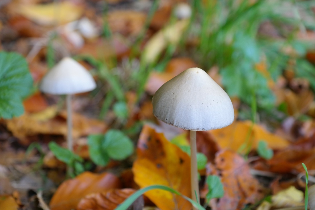 Psilocybe pelliculosa is a species of fungus in the family Hymenogastraceae. The fruit bodies, or mushrooms, have a conical brownish cap up to 2 cm (0.8 in) in diameter atop a slender stem up to 8 cm (3.1 in) long. It has a white partial veil that does not leave a ring on the stem. American mycologist Alexander H. Smith first described the species in 1937 as a member of the genus known today as Psathyrella; it was transferred to Psilocybe by Rolf Singer in 1958.
Psilocybe pelliculosa is found in the Pacific Northwest region of the United States and Canada, where it grows on the ground in groups or clusters along trails or forest roads in coniferous woods. A single collection has also been reported from Finland. The mushrooms contain the psychedelic compounds psilocybin and baeocystin, although at relatively low concentrations. Several mushroom species that are similar in appearance to P. pelliculosa can be distinguished by subtle differences in the form of the fruit body, or by microscopic characteristics.The cap of P. pelliculosa is initially sharply cone-shaped, and expands slightly over time to become broadly bell-shaped, but it never expands to become completely flat. The cap margin is pressed against the stem initially, and for a short time is appendiculate (has partial veil fragments hanging from the margin). The caps of mature specimens are smooth, sticky, and have translucent radial striations that reach dimensions of 0.8 to 2 cm (0.3 to 0.8 in) in diameter. The color ranges from umber to isabella (dark dingy yellow-brown) when the mushroom is moist, and changes to pinkish-buff when dry. The cap margin can have a greenish-gray tinge. The cap cuticle is a thin gelatinous covering that can be peeled off.[3] The gills have an adnate attachment to the cap, are narrow to moderately broad, closely spaced, and eventually separate from the stem. Young gills are cinnamon-brown in color, with lighter edges, but darken in maturity because they become covered with the dark spores. The stem is 6 to 8 cm (2.4 to 3.1 in) long by 1.5 to 2 mm (0.06 to 0.08 in) thick, and roughly equal in width throughout except for an slightly enlarged base. The lower region of the stem is brownish in color and has silky "hairs" pressed against the stem; the upper region is grayish and pruinose (lightly dusted with powdery white granules).[3] The flesh turns slightly bluish or greenish where it has been injured.[5] The application of a drop of dilute potassium hydroxide solution on the cap or flesh will cause a color change to pale to dark yellowish- to reddish-brown; a drop on the stem produces a less intense or no color change.[8] The spore print is purplish-brown.[11] Under the microscope, the spores appear dull purple-brown. They are ellipsoid to somewhat egg-shaped, and, according to Singer's original description, measure 8–10 by 4–5 μm.[3] A later study of specimens collected from British Columbia, Canada, instead reported a larger spore size range of 10–13 by 6–7 μm.[12] The spores have an apical germ pore.[5] The basidia (spore-bearing cells) are four-spored, hyaline (translucent), and measure 22–35 by 7–10 μm.[8] There are abundant cystidia that form a sterile band on the edges of the gills (cheilocystidia); these cystidia are smooth, inflated, and fusoid-ventricose (enlarged in the middle and tapered toward both ends) with an sharp tip, and measure 25–30 by 6–9 μm.[3] The cap cuticle (an ixocutis) is made of a layer of roughly horizontal, gelatinized, wavy, hyaline hyphae that are 0.8–5.5 μm in diameter. Wikipedia 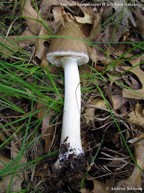 Amanita brunnescens[ Basidiomycetes > Agaricales > Amanitaceae > Amanita . . . ]
by Michael Kuo Sometimes called the “cleft-foot Amanita,” this eastern North American species features a variable, brown to whitish cap. The distinguishing feature of this relatively easily recognized mushroom is its stem, which, when acting the way it’s supposed to, ends in an abrupt basal bulb that is split or “chiseled” in one or more places, and discolors reddish brown. Unfortunately for would-be Amanita identifiers, however, Amanita brunnescens hasn’t read much Amanita literature and does not always manifest a perfectly bulbous, chiseled stem base; collections with wimpy, non-chiseled bases can be difficult to identify. Description: Ecology: Mycorrhizal with various hardwoods and conifers; growing alone, scattered, or gregariously; summer and fall; widely distributed and common east of the Rocky Mountains. Cap: 3-11 cm, convex, becoming broadly convex with a shallow central bump, or nearly flat; tacky at first or when wet; varying in color from grayish brown to whitish, often with a darker center; sometimes developing reddish brown stains; often somewhat streaked or mottled in appearance; usually featuring a few scattered, randomly distributed white to grayish or tan warts; the margin often becoming faintly lined for a few mm. Gills: Free from the stem; white; close or crowded; not discoloring, or sometimes discoloring brownish. Stem: 6-12 cm long; 0.5-1.5 cm thick above the bulb; tapering to apex; bald or silky; with a relatively persistent, skirtlike, white ring that sometimes develops a reddish brown edge and often collapses against the stem; usually ending in an abrupt, rimmed basal bulb that is “chiseled” or split vertically in one or more places; discoloring and bruising reddish brown, especially near the base; volval remnants usually absent but occasionally present as a few patches along the upper rim of the bulb. Flesh: White throughout; firm; not discoloring, or sometimes discoloring or bruising reddish brown, especially around worm channels. Odor: Not distinctive. Chemical Reactions: KOH on cap surface negative; on flesh in stem base slowly slightly yellowish. Spore Print: White. Microscopic Features: Spores 6.5-10 µ; smooth; globose or subglobose; amyloid. Basidia without basal clamps; 4-spored. Pileipellis a cutis or ixocutis of hyphae 2-6 µ wide. Lamellar trama bilateral; subhymenium ramose or with inflated cells. REFERENCES: Atkinson, 1918. (Smith, 1949; Smith, Smith & Weber, 1979; Arora, 1986; Jenkins, 1986; Phillips, 1991/2005; Lincoff, 1992; Metzler & Metzler, 1992; Barron, 1999; Roody, 2003; McNeil, 2006; Miller & Miller, 2006; Binion et al., 2008; Tulloss, cont. upd..) Herb. Kuo 06229501, 07200203, 07200204, 07200205, 06120301, 07160703, 07100802, 10170904, 06221010. Amanita brunnescens var. pallida, recognized by some authors, is virtually identical, but has a nearly white cap. I have collected it growing alongside the typical, brown variety, and I have seen specimens that seemed to “intergrade” between the colors of the putative varieties; for this reason I am treating var. pallida as a synonym of the type variety. Further Online Information: Amanita brunnescens at Studies in the Amanitaceae Amanita brunnescens at Roger’s Mushrooms Kuo, M. (2013, March). Amanita brunnescens. Retrieved from the MushroomExpert.Com Web site: http://www.mushroomexpert.com/amanita_brunnescens.html 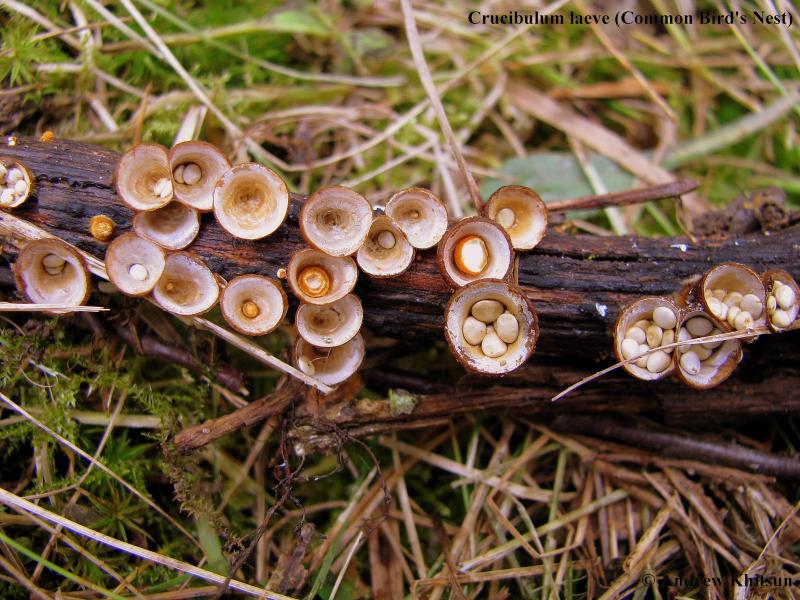 Crucibulum laeve
[ Basidiomycota > Agaricales > Agaricaceae > Crucibulum . . . ] by Michael Kuo Crucibulum laeve is probably the most common and frequently encountered bird’s nest fungus in temperate North America. Three features serve to identify it successfully: its yellowish colors; the “lid” over the nest (in young specimens), covering the eggs; and the tiny cords that attach the eggs to the nest. Observing this last feature requires some patience with a very tiny tool–say, a needle or a pin–and a hand lens. The cord, which is called a “funiculus” in Mycologese, is the egg’s mechanism for attaching itself to sticks, leaves, and other plant debris. When a raindrop falls into the nest, the eggs are projected out of the cup. As this happens, the cord is stretched to its limit–then breaks away from the nest, remaining attached to the egg. Where the cord was attached to the nest, it becomes frayed, since it was torn away. The little frayed ends are adhesive, and when they come into contact with, for example, a leaf, they attach themselves. This stops the flight of the egg, which then swings back and attaches itself to the leaf as well . . . rather like what would happen to a kite if you were to let it sail away after coating your end of the string with glue. Description: Ecology: Saprobic; growing alone, scattered, or densely gregariously on woodland debris (sticks, leaves, nutshells, needles, etc.), woodchips, old furniture, dung, and so on (but not typically on the ground alone or on larger logs); spring through fall (or in winter in warmer climates); widely distributed in North America. Nest: 2-9 mm high; 4-10 mm across; at first cushion-shaped to round, and closed by a mustard yellow to dull yellow “lid”; later becoming cup-shaped or goblet-shaped, the lid disappearing; outer surface yellowish at first, remaining yellow or darkening to nearly brown, velvety or fairly bald; inner surface bald and shiny, whitish to grayish. Eggs: To 2 mm wide; shaped like flattened circles or ellipses; tough; attached to the nest by tiny cords; pale tan to buff. Microscopic Features: Spores 6-9 x 3-5 µ; smooth; elliptical. REFERENCES: (Hudson, 1778) Kambly, 1936. (Saccardo, 1888; Brodie, 1975; Smith, Smith & Weber, 1981; Arora, 1986; States, 1990; Phillips, 1991/2005; Lincoff, 1992; Horn, Kay & Abel, 1993; Evenson, 1997; Barron, 1999; Roody, 2003; McNeil, 2006; Miller & Miller, 2006.) Herb. Kuo 06079601, 06300703, 06080801, 06121302 Kuo, M. (2014, February). Crucibulum laeve. Retrieved from the MushroomExpert.Com Web site: http://www.mushroomexpert.com/crucibulum_laeve.html 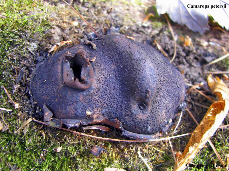 [Ascomycetes > Boliniales > Boliniaceae > Camarops . . . ]by Michael Kuo
Have you ever seen those ridiculous “faces” that people put on trees in their yards? This mushroom represents what would happen if the Addams Family got hold of the idea. It’s creepy and its kooky, mysterious and spooky. It’s altogether ooky . . . Camarops petersii. For more of Uncle Fester’s lawn decorations, see Pisolithus tinctorius and Scleroderma polyrhizum. Camarops petersii looks like a bulging black eye, complete with eyelids, stuck to the side of a dead log. The eyelid is a veil that protects the young mushroom but soon ruptures to expose the spore-producing surface. The range of Camarops petersii in North America extends from eastern North America to at least Kansas and Cuba. Description: Ecology: Saprobic on the decorticated wood of fallen oaks (early records for the species also included wood of the now extinct American chestnut as a substrate); growing alone or in small clusters; late summer and fall; eastern North America to Kansas and Cuba. Fruiting Body: 2-7 cm wide; up to about 2 cm high; sub-circular or broadly elliptical in outline; cushion-shaped, with a somewhat narrowed base; upper surface black and shiny, covered with pimple-like dots (and covered with black slime when the mushroom is producing spores); encased in a black, feltlike veil that soon ruptures and becomes a sheath around the sides of the fruiting body, with a ragged upper edge; interior tough and brownish, filled with black channels and pockets but not featuring concentric zones. Chemical Reactions: Tissues from dried specimens brownish when crushed in KOH. Microscopic Features: Asci 8-spored; deliquescing along with the paraphyses so that spores are exuded from the perithecia in a gelatinous black matrix (according to Nannfeldt [1972], “[t]he ascus has lost its gun function”). Spores 6-8.5 x 3-4.5 µ smooth; broadly elliptical at one end and broadly fusiform at the other; with a tiny pore at the narrowed end; usually biguttulate in KOH or water mounts; purplish gray in KOH. REFERENCES: (Berkeley & Curtis, 1868) Nannfeldt, 1972. (Saccardo, 1882; Nannfeldt, 1972; Horn, Kay & Abel, 1993.) Herb. Kuo 10160407, 09190602. Kuo, M. (2007, February). Camarops petersii. Retrieved from the MushroomExpert.Com Web site: http://www.mushroomexpert.com/camarops_petersii.html 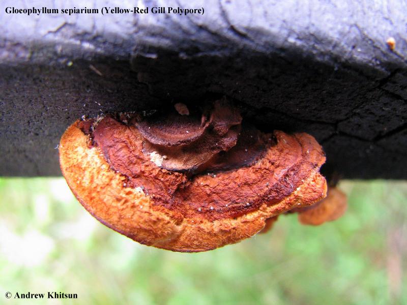 Gloeophyllum sepiarium
[ Basidiomycetes > Polyporales > Gloeophyllaceae > Gloeophyllum . . . ] by Michael Kuo Decomposing the deadwood of conifers across North America, Gloeophyllum sepiarium is fairly easily recognized. When fresh and very young its cap is more or less orange, but as it matures brown colors replace the orange from the center outwards. The underside of this polypore features gills–which is a bit odd, since polypores have pores rather than gills! (For help sorting through this mess, see the essay on the page for Lenzites betulina, another “gilled polypore.”) Other distinguishing features include the brown flesh and the black reaction to KOH, as well as microscopic features. Description: Ecology: Saprobic on the deadwood of conifers and, occasionally, hardwoods (especially those, like quaking aspen, that grow in conifer-dominated ecosystems); causing a brown rot; growing alone or gregariously; appearing in woods, but not infrequently found on lumber in urban settings; annual or reviving to be perennial; summer and fall (and over winter in warm climates); widely distributed in North America. Cap: Single or compound (and then either fused laterally or with loosely arranged lobes arising from a central point); up to about 12 cm across and 8 cm deep; semicircular, irregularly bracket-shaped, or kidney-shaped; flattened-convex; velvety to hairy; rugged; with concentric zones of texture and color; at first yellow to orange, becoming yellow-brown to dark brown or nearly black toward the point of attachment but usually remaining yellow to orange on the growing margin. Gills: Irregular and often fusing; fairly close; often mixed with slot-like pores; edges yellow-brown becoming darker brown with age; faces creamy to pale brownish, darkening with age; up to about 1 cm deep. Stem: Absent Flesh: Dark rusty brown or dark yellow-brown; corky. Chemical Reactions: KOH black on flesh. Spore Print: White. Kuo, M. (2010, February). Gloeophyllum sepiarium. Retrieved from the MushroomExpert.Com Web site: http://www.mushroomexpert.com/gloeophyllum_sepiarium.html
6/1/2015
June 2015 Boletus subtomentosus (Suede, Yellow-Cracked Bolete), synonym Xerocomus subtomentosus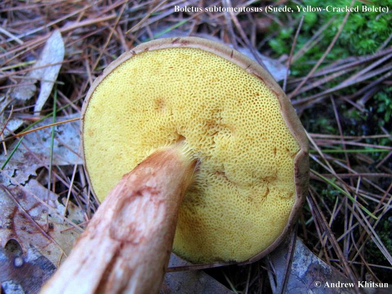 Boletus subtomentosus
[ Basidiomycetes > Boletales > Boletaceae > Boletus . . . ] by Michael Kuo This common bolete can be a nightmare to identify if you don’t have a drop of common household ammonia handy. Maybe it’s just me, but every time I find Boletus subtomentosus, I promptly forget most of what I know about boletes and spend hours trying to figure out what I’ve found. It’s just such an average bolete; nothing really seems to stand out as a distinctive identification feature. Except the ammonia thing: a drop on the cap produces an instant mahogany red reaction. This will separate Boletus subtomentosus from some of the other species of “boletish” (the ones that are velvety-ish and brownish-capped, with yellowish pore surfaces that bruise-ish faintly bluish or greenish), including Boletus spadiceus and Boletus illudens, both of which flash green with ammonia before resolving to reddish brown. Description: Ecology: Mycorrhizal with hardwoods or conifers; growing alone or scattered; summer and fall; widely distributed in North America. Cap: 5-18 cm; convex, becoming broadly convex or almost flat; dry; finely velvety; often becoming cracked in age, with yellowish flesh showing in the cracks; olive to olive brown, or yellowish brown, sometimes faintly reddish in age; margin incurved when young, with a projecting sterile portion. Pore Surface: Yellowish; bruising slowly greenish, then brown; 1-2 pores per mm; tubes 1-2.5 cm deep. Stem: 4-10 cm long; 1-2 cm thick; more or less equal, or tapering near the base; solid; yellowish, with reddish brown stains; basal mycelium sulphur yellow; not reticulate, but often with ridges approaching an obscure reticulum, especially near the apex; bruising slowly brownish to reddish brown on handling. Flesh: White or pale yellow; not staining on exposure, or staining faintly blue. Odor and Taste: Not distinctive. Chemical Reactions: Cap instantly mahogany red with ammonia. Spore Print: Olive brown. Kuo, M. (2003, March). Boletus subtomentosus. Retrieved from the MushroomExpert.Com Web site: http://www.mushroomexpert.com/boletus_subtomentosus.html |
 RSS Feed
|
If You Suspect a Poisoning
If you suspect you have consumed a poisonous mushroom, contact a physician, the closest hospital ER, poison control center, or dial 911, depending on the severity of the reaction. US Poison Control: 1-800-222-1222 The North American Mycological Association (NAMA) has information that may also be of help. Click here. We do not ID mushrooms through this website.
If you are in need of an ID consider uploading quality photos with multiple views of your specimen and descriptions of your find to Mushroom Observer or iNaturalist including our projects or post in Wild Food Wisconsin or Mushroom Identification Group. If you contact us and provide a way to get back to you, we may be able to provide suggestions for more identification resources you can use. You are always responsible for your own decisions taken on the basis of identification resources. |
Wisconsin Mycological Society
Wisconsin Mycological Society (WMS) is dedicated to the study and enjoyment of mushrooms and other fungi throughout the state of Wisconsin. Education, safety, sustainability, community, and connecting with nature are our goals.
|